Electrons - Photons - Neurons
Biological processes are generally based on the changes and activities on the molecular and cellular level. Here, they interact in manifold ways with the surrounding tissue. Thus, in order to understand such biological processes they need to be studied where they take place – within the living tissue. The necessary high-resolution observations are only possible with the aid of optical microscopes. Although the light-microscopy is known since the early 17th century and is an integral part of scientific research, the technique is far from being outmoded. Especially the development of fluorescent dyes has made the microscopy to one of the most important technologies of today's biomedical research. With the aid of these dyes, individual cells, parts of specific cells or specific cellular processes become visible under the microscope.
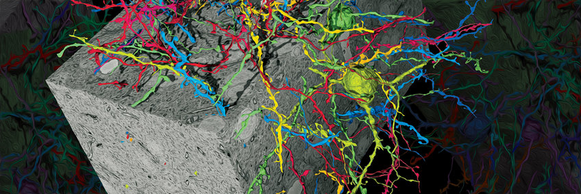
The aim of our research is the development of new and better optical methods for the biomedical research. One of our successes is the co-development of the Multiple-Photon-Excitation-Microscope. This microscope reduces the strong diffusion of light, which is especially prominent in brain tissue, and which limits the use of conventional light microscopes. A further success is the development of the Serial Block Face Scanning Electron Microscopy: In an automated process, an electron microscope scans the surface of a tissue sample and saves the resulting picture. Next, the device removes an ultra-thin tissue slice and again scans the now novel surface of the sample. In this way, a tissue sample is scanned level for level before it is reassembled at the computer into a 3D image of the original sample. This is an optimal starting point to investigate the network structure and individual nerve cell connections of the brain.